融合光谱与激光散斑技术,实现烧伤深度的快速定量评估
Multimodal Optical Imaging Platform for Quantitative Burn Assessment
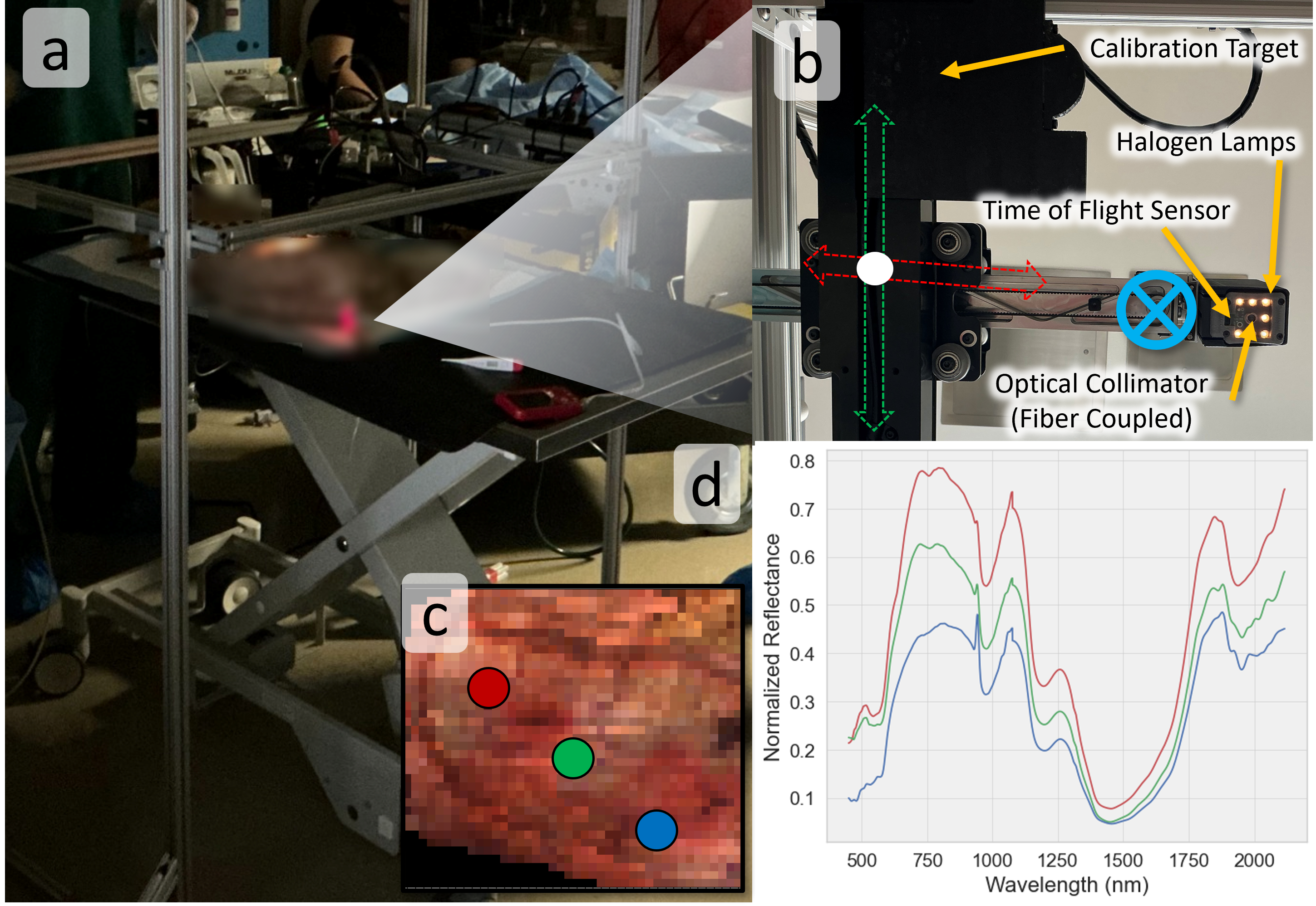
- 结合可见-短波红外光谱与激光散斑,同时分析组织生化成分和血流灌注
- 在1000nm以上波段发现水、脂质、胶原吸收特征,提升烧伤组织辨识度
- 无需标注数据即可自动提取特征,适合战场或救援现场快速部署
烧伤严重程度的准确评估在伤后初期仍面临重大临床挑战,主要因缺乏客观检测深层组织损伤的方法。这一局限在战场或大规模伤亡场景中尤为关键,快速可靠的烧伤深度评估对分诊和手术决策至关重要。本文提出一种多模态光学成像框架,为紧凑、低尺寸、重量和功耗(低SWaP)的野外可部署设备奠定基础。系统整合宽带高光谱成像(VSWIR,400–2100 nm)与激光散斑对比成像,联合评估生物化学组成与微血管灌注情况。利用短波红外(SWIR,>1000 nm)波段,开发并验证了与水、脂质、胶原吸收特征相关的新型深层组织参数,显著增强烧伤组织区分能力及严重程度分类效果。通过无监督学习方法实现光谱特征提取、波段降维与聚类,并基于组织病理学验证,为严苛环境下早期定量烧伤评估提供坚固、数据驱动的技术基础。
原文摘要 · Abstract (English)
Accurate assessment of burn severity at injury onset remains a major clinical challenge due to the lack of objective methods for detecting subsurface tissue damage. This limitation is critical in battlefield and mass-casualty settings, where rapid and reliable evaluation of burn depth is essential for triage and surgical decision-making. We present a multimodal optical imaging framework that establishes the foundation for a compact, low-size, weight, and power (low-SWaP) field-deployable device for quantitative burn assessment. The system integrates broadband hyperspectral imaging (VSWIR, 400 -- 2100 nm) with laser speckle contrast imaging to jointly evaluate biochemical composition and microvascular perfusion. Using short-wave infrared (SWIR, >1000 nm) wavelengths, we developed and validated novel deep-tissue parameters linked to water, lipid, and collagen absorption features that enhance burn-tissue separability and burn severity classification. We implemented and validated unsupervised learning methods for spectral feature extraction, band down-selection, and clustering against histology, establishing a foundation for a rugged, data-driven device for early quantitative burn evaluation in austere environments.
Thank you to arXiv for use of its open access interoperability. PaperDance 不是 arXiv 官方产品;中文卡片由大模型生成,请以原文为准。